PINGUECULA(E)
Pingueculae are yellow-white, flat, or slightly raised conjunctival lesion(s), usually in the interpalpebral fissure adjacent to the limbus, but not involving the cornea.
Elastotic degeneration develops in the deep conjunctival layers upon chronic exposure to sunlight and irritation, e.g. from wind and dust. They commonly occur in people in sunny climates.
PTERYGIUM/PTERYGIA
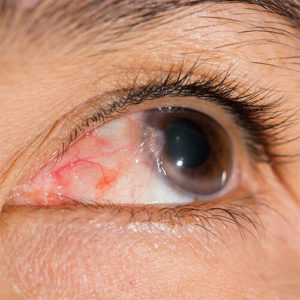 Pterygia are triangular lesions of the bulbar conjunctiva that extend onto the cornea. They enlarge in response to sunlight exposure and environmental irritants such as wind and dust.
They most often develop from pingueculae, which are elevated, yellow-white, and sometimes injected, round or amorphous lesions that appear on either side of the cornea.
CONSERVATIVE TREATMENT
- UV protection with sunglasses and a hat while outdoors.
- Artificial Tears can be used as needed for comfort.
- Over-the-counter medications such as Naphcon A may be instilled up to QID.
- Steroid eye drops such as FML BID or prednisolone acetate QID may bring additional relief.
- Avoid windy, highly ventilated, smoky environments, and contact lenses if they are irritating.
SURGICAL EXCISION
Pingueculae are infrequently excised. However, when the patient experiences excessive irritation refractory to topical medications, the lesion interferes with contact lens wear, it is excessively large and/or red, or it has progressed into a bothersome pterygium, then removal can bring the patient relief from symptoms.
When pterygia induce significant astigmatism (by pulling on the cornea), jeopardize the visual axis, or cause foreign body sensation, burning, or redness inadequately responsive to topical medication(s), excision may be considered.
Dr. Shin excises the lesions in an outpatient surgery center. Both a pinguecula and a pterygium are peeled from the underlying layers of the eye and blood vessels are cauterized.
In the case of a pinguecula, an amniotic membrane graft is placed over the area where the pinguecula was removed and is affixed to the eye with tissue glue to prevent the area from becoming dry and inflamed.
Dr. Shin tailors her surgical approach for patients with pterygium and an increased risk of recurrence, for example, for golfers and landscapers; She may include dilute Mitomycin C to inhibit regrowth and/or transfer a piece of the patient’s own surface tissue from another location on the same eye (conjunctival autograft) instead of placing an amniotic membrane over the scleral bed.