DERMOID/EPIDERMOID CYSTS
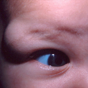 In children, these are commonly smooth, palpable, painless, mobile or non-mobile, oval, benign masses that slowly enlarge near the lateral brow and upper lid adjacent to the zygomaticofrontal suture.
Rarely they may be located at the nasofrontal suture. If the mass is located at the nasal aspect of the brow, the possibility of an encephalocele should be considered.
Dermoid cysts develop from dermal elements and epidermal cysts form from epidermal elements that get pinched off along suture lines during embryonic development.
The cyst may erode through or cause a deformity in the bone in or surrounding the orbit and if it ruptures, it can cause an acute inflammatory response within the eyelid or orbit.
If excised, they should be removed in their entirety and sent to a lab for pathological analysis.
LIPODERMOID OR DERMOLIPOMA
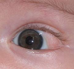 These are congenital, pale yellowish-white, soft, fluctuant, fatty benign tumors located subconjunctivally, most often in the super-temporal quadrant of the orbit and most easily observed in inferonasal gaze.
The lesion is fixed to the conjunctiva, which is markedly thickened over it and may contain fine hairs, sweat, sebaceous glands, and hair follicles. They are frequently associated with Goldenhar syndrome.
The borders are often ill-defined, and the tumor may be tightly fused with the levator aponeurosis, the lacrimal gland, the lateral rectus muscle, and less commonly, the peripheral cornea, all of which make removal difficult.
This type of lesion is most commonly just observed, but when it is visible between the eyelids or causes difficulty with outward rotation of the eye because of its bulk, partial resection can be performed.
DACRYOCYSTOCELE
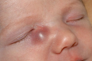 This enlargement of the lacrimal sac results when amniotic fluid or mucus (produced by the goblet cells of the lacrimal sac) becomes trapped within the tear sac in conjunction with a nasolacrimal duct obstruction.
It is initially sterile, but an infection may develop within or around the tear sac. Congenital swelling above the medial canthal tendon, especially in the midline, may represent an encephalocele.
It may respond to massage and topical antibiotics, but if it does not resolve in 1-2 weeks or an infection develops, nasolacrimal duct probing is necessary.